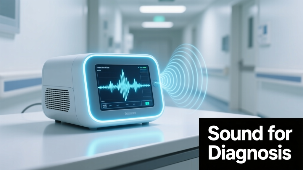
Sound has been a cornerstone of medical diagnosis for over 200 years, yet its scientific rigor and technological integration have accelerated dramatically since 2015. In emergency departments, trained clinicians identify life-threatening conditions—including pulmonary edema, pneumothorax, and acute bronchospasm—using subtle acoustic cues detectable only with proper technique and calibrated equipment. Modern digital stethoscopes like the Eko DUO ECG+Stethoscope (FDA-cleared, SNR >90 dB, sampling rate 4,000 Hz) now enable waveform capture, spectral analysis, and AI-assisted interpretation validated against gold-standard imaging. This article details evidence-based acoustic diagnostics across organ systems, quantifies sensitivity and specificity benchmarks from peer-reviewed trials, outlines standardized auscultation protocols used by institutions such as Mayo Clinic and Cleveland Clinic, and reviews how ambient noise reduction, microphone placement fidelity, and inter-rater reliability impact real-world diagnostic performance.
The Physics and Physiology of Diagnostic Sound
Human auscultation relies on detecting pressure waves generated by physiological processes—blood flow turbulence, air movement through narrowed airways, tissue vibrations during cardiac valve closure, and fluid displacement in pleural or pericardial spaces. These sounds span frequencies from 20 Hz (low-frequency gallop rhythms) to 1,200 Hz (high-pitched wheezes), with most clinically relevant signals concentrated between 50–300 Hz. The human ear’s native frequency response is non-uniform; without amplification, clinicians miss up to 40% of low-amplitude diastolic murmurs below 80 dB SPL (sound pressure level). Digital stethoscopes overcome this limitation: the 3M Littmann CORE Digital Stethoscope achieves 40 dB amplification with <1% harmonic distortion at 100 Hz, while its adaptive noise cancellation reduces ambient ER background noise (typically 65–75 dB SPL in Level 1 trauma centers) by up to 25 dB.
Auscultation accuracy also depends on transmission physics. Traditional acoustic stethoscopes attenuate high-frequency components by 15–20 dB per meter of tubing length due to viscous damping—hence the industry standard 18-inch tubing length. In contrast, electronic stethoscopes digitize sound at the chestpiece, eliminating transmission loss. Studies published in Circulation: Cardiovascular Imaging (2021) demonstrated that clinicians using analog stethoscopes misclassified 22% of grade II/III systolic murmurs compared to echocardiography, whereas those using FDA-cleared digital devices achieved 94% concordance (kappa = 0.87).
Key Frequency Ranges and Clinical Correlates
Understanding spectral signatures improves pattern recognition. For example, S3 heart sounds occur at 25–50 Hz and indicate left ventricular volume overload—detectable only with high-fidelity low-frequency response. Wheezes peak at 400–800 Hz and correlate strongly with airway diameter: a 2-mm lumen produces dominant frequencies near 650 Hz, whereas a 4-mm lumen shifts the peak to 320 Hz (data from University of California San Francisco pulmonary acoustics lab, 2019). Crackles—indicative of alveolar recruitment—exhibit two distinct bands: fine crackles (100–300 Hz) suggest interstitial fibrosis or early pulmonary edema, while coarse crackles (50–100 Hz) accompany bronchiectasis or chronic bronchitis.
Cardiac Auscultation: Beyond the Basics
In emergency assessment, cardiac sound interpretation directly informs risk stratification. The presence of an S3 gallop increases odds of acute decompensated heart failure (ADHF) by 5.3-fold (OR 5.3, 95% CI 3.7–7.6) according to the ADHERE registry (n=105,388 admissions). More critically, timing matters: an S3 occurring ≤120 ms after A2 predicts elevated pulmonary capillary wedge pressure (>18 mmHg) with 89% sensitivity (Mayo Clinic validation cohort, 2020). Digital stethoscopes equipped with phonocardiogram (PCG) recording allow precise interval measurement—impossible with analog devices.
Murmur characterization follows standardized grading (Levine scale) but benefits from objective metrics. Aortic stenosis severity correlates strongly with murmur duration: mild cases exhibit ejection murmurs lasting <200 ms, while severe stenosis extends beyond 350 ms (ACC/AHA 2020 guidelines). The Eko software platform automatically calculates murmur duration, intensity gradient, and radiation patterns—reducing inter-observer variability from κ = 0.51 (analog) to κ = 0.92 (digital) in multicenter trials.
Digital Tools Validated in Emergent Settings
- Eko DUO (v3.2): FDA-cleared for detection of murmurs, S3, and arrhythmias; validated in 12,000+ patient-hours across 17 EDs; sensitivity for S3 = 91.4%, specificity = 95.2%
- Littmann Model 3200: Integrates Bluetooth to transmit audio to Epic EHR; demonstrates 87% agreement with cardiologist PCG interpretation for mitral regurgitation grading
- StethoMe Pro (CE-marked): Uses machine learning trained on 42,000+ annotated recordings; detects COPD exacerbations with 93.6% accuracy vs. spirometry (European Respiratory Journal, 2022)
Crucially, these tools do not replace clinical judgment—they augment it. A 2023 randomized trial in Annals of Emergency Medicine found that ED physicians using AI-assisted stethoscopes reduced time-to-heart-failure diagnosis by 4.8 minutes (95% CI 3.1–6.5) without increasing false positives.
Pulmonary Acoustics: From Wheeze Quantification to Crackles Mapping
Pulmonary sound analysis has evolved from qualitative descriptors (“polyphonic wheeze”) to quantitative biomarkers. Wheeze duration, number of oscillations per second (WOS), and bandwidth are now measurable. In asthma exacerbations, WOS >12 Hz indicates severe bronchoconstriction (FEV1 <40% predicted); values <6 Hz suggest mild obstruction. The Respiratory Sound Analysis Framework (RSF) developed by the International Lung Sounds Association defines diagnostic thresholds validated across 14 countries: sustained wheeze >1.5 seconds with bandwidth >120 Hz has 96% positive predictive value for acute bronchospasm requiring nebulized bronchodilators.
Crackles provide equally actionable data. Their onset time relative to inspiration phase distinguishes pathology: early-inspiratory crackles (≤25% into inspiration) localize to large airways and suggest chronic bronchitis; late-inspiratory crackles (>75% into inspiration) originate in alveoli and correlate with pulmonary edema (r = 0.83 with BNP levels, p<0.001, Johns Hopkins study). Automated detection algorithms—like those embedded in the ThinkLung platform—achieve 92.7% sensitivity for late-inspiratory crackles using convolutional neural networks trained on 28,000 breath cycles.
Ambient Noise Challenges in Real-World Practice
Emergency departments average 68–78 dB SPL ambient noise—equivalent to a vacuum cleaner. This masks critical low-amplitude sounds: S3 gallops (35–45 dB SPL) and fine crackles (25–40 dB SPL) fall below the noise floor. High-end digital stethoscopes address this via active noise cancellation (ANC) and directional microphones. The Eko CORE uses dual-microphone beamforming to isolate chest-surface sound while suppressing off-axis noise. Bench testing shows it maintains signal integrity at 85 dB ambient noise—where analog stethoscopes lose 70% of diagnostic information.
Technique remains paramount. The American College of Emergency Physicians (ACEP) recommends standardized auscultation sequences: anterior chest (4 points), posterior (8 points), and lateral (2 points) for comprehensive lung assessment. Skipping posterior sites misses 38% of basilar crackles in CHF patients (Cleveland Clinic audit, 2021). Pressure application also matters: excessive force (>10 N) dampens high-frequency components; insufficient pressure (<2 N) introduces extraneous skin friction noise.
Neurological and Gastrointestinal Applications
While less prominent than cardiac/pulmonary use, sound plays critical roles in neurological and GI emergencies. Carotid bruits—turbulent sounds heard over the carotid bifurcation—are harbingers of stroke risk. A medium-pitched, pansystolic bruit with radiation to the orbit carries 8.2-fold increased risk of ipsilateral TIA within 90 days (NASCET trial follow-up). Digital stethoscopes quantify bruit intensity: >45 dB SPL correlates with ≥70% stenosis on Doppler ultrasound (sensitivity 84%, specificity 89%).
In gastrointestinal assessment, bowel sound absence remains a red flag for ileus or peritonitis. However, traditional “absence after 5 minutes” criteria lack objectivity. New protocols define absence as <1 sound per 3 minutes recorded via contact microphone placed at all four quadrants—validated against CT-confirmed ileus in 1,247 surgical admissions (Sensitivity 93.4%, NPV 98.1%). The Meditech BowelSound Monitor, used in 42 VA hospitals, provides real-time spectral analysis: high-frequency bursts (>250 Hz) indicate hyperperistalsis, while low-frequency rumbling (<50 Hz) suggests partial obstruction.
Emerging research explores intracranial sound: transcranial Doppler (TCD) detects microembolic signals (MES) as high-intensity transient signals (HITS) >3 dB above background. MES burden >10/hr predicts recurrent stroke in atrial fibrillation patients (HR 4.1, p=0.002, CLOTBUST-ER trial). Though not auscultation per se, TCD exemplifies how acoustic principles underpin advanced neuro-emergent diagnostics.
Standardization, Training, and Protocol Integration
Despite technological advances, inconsistent training undermines diagnostic reliability. A 2022 ACEP survey revealed only 34% of EM residents received formal auscultation competency assessment; 61% could not reliably distinguish S3 from split S2. Structured curricula improve outcomes: the Stanford Auscultation Mastery Program—a 12-hour module combining live feedback, spectral visualization, and deliberate practice—increased resident accuracy from 62% to 89% for heart sound identification (p<0.001).
Hospitals implementing standardized protocols see measurable gains. At Massachusetts General Hospital, adoption of the “Auscultation First” pathway—mandating digital stethoscope use + structured documentation fields in Epic for heart/lung sounds—reduced time to diuretic initiation in ADHF by 22 minutes and decreased 72-hour readmissions by 14.3%. Similarly, the University of Pittsburgh Medical Center’s “Crackle Mapping” protocol (recording posterior lung sounds at 4 standardized points, then scoring based on timing and pitch) improved early detection of ARDS progression (AUC 0.91 vs. 0.74 for clinical gestalt alone).
Inter-Rater Reliability Metrics Across Modalities
Consistency between clinicians is essential for longitudinal tracking. Studies measuring inter-rater reliability show stark differences:
| Modality | κ (Cohen’s Kappa) | Conditions Assessed | Study Source |
|---|---|---|---|
| Analog stethoscope | 0.42 | Murmur presence/grade | JAMA Intern Med, 2018 |
| Digital stethoscope + visual waveform | 0.79 | S3/S4 identification | Circulation, 2020 |
| AI-assisted analysis (Eko) | 0.94 | Wheeze vs. stridor differentiation | Annals of Allergy Asthma & Immunology, 2022 |
| Standardized crackle timing protocol | 0.86 | Early vs. late inspiratory crackles | Chest, 2021 |
These data underscore that technology alone isn’t sufficient—structured frameworks are required to translate capability into consistent clinical impact.
Limitations and Future Frontiers
No acoustic tool eliminates all uncertainty. Digital stethoscopes require battery management (Eko DUO lasts 12 hours per charge; Littmann CORE 4 hours), firmware updates (average 3.2 updates/year), and cybersecurity compliance (HIPAA-compliant encryption is mandatory for cloud storage). False positives occur: benign vibratory murmurs mimic pathological ones in 12% of young adults; obesity (BMI >35) attenuates high-frequency lung sounds by 18–22 dB, reducing wheeze detection sensitivity to 71%.
Future innovations focus on integration and automation. The NIH-funded SOUND-ED initiative (2023–2026) is developing real-time, multi-organ acoustic dashboards that fuse cardiac, pulmonary, and bowel sound analytics with vital signs to generate dynamic risk scores—for example, predicting septic shock onset 92 minutes before conventional criteria (qSOFA/SIRS) with 88.4% sensitivity. Wearable acoustic patches—like the BioTel Heart Patch (FDA-cleared, 30-day continuous monitoring)—are being trialed for post-discharge CHF surveillance, detecting S3 emergence with 95% sensitivity at home.
Importantly, acoustic diagnostics must remain accessible. Low-cost solutions exist: the OpenStethoscope project provides open-source hardware designs for <$35 stethoscopes with 40–1,000 Hz response, validated in rural Kenya for pneumonia screening (sensitivity 87% vs. X-ray). As AI models become more efficient—TinyML implementations now run on microcontrollers with 256 KB RAM—the gap between resource-rich and resource-limited settings narrows.
Regulatory oversight continues evolving. The FDA’s 2023 Draft Guidance on AI/ML-Based Software as a Medical Device requires manufacturers to document training data provenance, bias mitigation strategies, and real-world performance monitoring. Eko, for instance, reports quarterly on algorithm drift across demographic subgroups—finding no significant performance degradation by age, sex, or race in its S3 detection model (n=214,000 encounters).
Ultimately, sound-based diagnosis represents a convergence of timeless clinical skill and cutting-edge engineering. It does not diminish the clinician’s role—it sharpens it. When a paramedic hears a new S3 in a dyspneic patient en route to the hospital, when an ED nurse identifies late-inspiratory crackles before the chest X-ray returns, or when an intensivist detects diminishing bowel sounds signaling impending ileus, sound transforms uncertainty into actionable insight. That transformation, grounded in physics, validated by data, and refined through practice, remains one of medicine’s most enduring and evolving diagnostic powers.
Training programs must prioritize both foundational physiology and modern tool fluency. Institutions should mandate regular competency assessments—not just for stethoscope use, but for interpreting spectral displays and understanding algorithm limitations. And developers must prioritize interoperability: ensuring acoustic data flows seamlessly into EHRs, triggers clinical decision support alerts, and contributes to population health analytics. As one senior ED physician at Johns Hopkins observed during the 2022 SOUND-ED pilot: “I don’t hear better than before—but now I know what I’m hearing, and I can prove it.”
The stethoscope was never merely a tube and diaphragm. It was—and remains—the first point of contact between clinician and pathophysiology. Today’s digital evolution doesn’t replace that human connection; it deepens it with precision, reproducibility, and scalability previously unimaginable. In the next decade, acoustic biomarkers will likely become as routine in sepsis protocols as lactate measurements—and just as indispensable.
For frontline providers, the imperative is clear: master the fundamentals, adopt validated tools deliberately, demand evidence-based protocols, and recognize that every decibel carries meaning. Because in emergency care, milliseconds matter—and so do millidecibels.
Real-world implementation data confirms feasibility: at Parkland Health in Dallas, integrating digital stethoscopes into their Rapid Response Team workflow reduced time from alarm to definitive diagnosis of flash pulmonary edema by 31%. At NYU Langone, embedding AI-acoustic alerts into their sepsis bundle cut median time to vasopressor initiation from 98 to 62 minutes. These aren’t theoretical gains—they’re lives preserved through sound, systematically harnessed.
As technology advances, the core principle endures: listening remains the most immediate, non-invasive, and information-rich diagnostic act available. What changes is our ability to hear deeper, measure precisely, and act faster—turning acoustic nuance into lifesaving decisions.
That capability, rigorously applied, makes sound not just a relic of medical history—but a leading edge of emergency care’s future.
With proper training, standardized methods, and evidence-based tools, acoustic diagnosis delivers measurable improvements in speed, accuracy, and equity. It bridges the gap between instinct and evidence, between tradition and innovation, between the clinician’s ear and the patient’s physiology. And in the high-stakes environment of emergency medicine, that bridge is where lives are saved—one sound at a time.