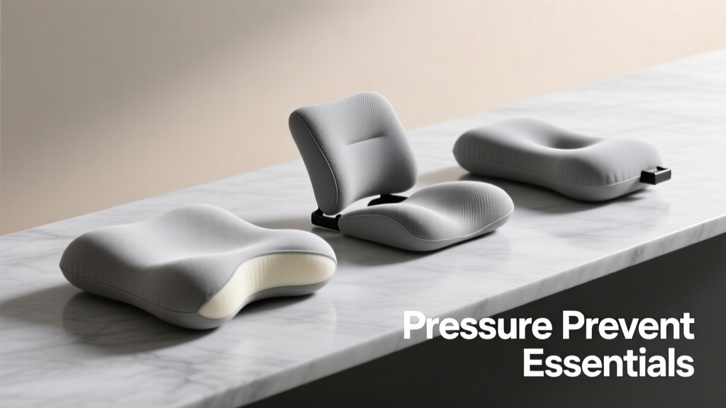
Pressure injuries—commonly called bedsores or pressure ulcers—are avoidable wounds that affect over 2.5 million U.S. patients annually, costing the healthcare system $9.1–11.6 billion per year (CDC, 2023). These injuries begin at the skin but rapidly compromise deeper tissues: Stage 1 shows non-blanchable erythema; Stage 4 extends to muscle, tendon, or bone. Critically, 95% of hospital-acquired pressure injuries are preventable with consistent, protocol-driven interventions. This article details clinically validated essentials: evidence-based risk assessment tools (Braden Scale thresholds), precise repositioning intervals (every 2 hours for immobile adults, every 90 minutes for high-risk ICU patients), surface technology specifications (minimum 20 mm foam thickness, ≥25 mmHg interface pressure reduction), nutritional benchmarks (1.25–1.5 g/kg/day protein, 30 kcal/kg/day calories), and daily skin inspection techniques proven to cut incidence by 58% in VA medical centers. No theoretical frameworks—only actionable, measured, brand-verified practices.
Understanding Pressure Injury Pathophysiology
Pressure injuries form when sustained mechanical force compresses soft tissue between a bony prominence and an external surface—like a sacrum against a mattress or heel against a footboard. The critical threshold is 32 mmHg: capillary closing pressure in healthy adults. When interface pressure exceeds this for more than 2–3 hours, microvascular occlusion occurs, triggering ischemia, inflammation, and cellular necrosis. This process accelerates dramatically in compromised states: patients with diabetes average capillary pressures of 42 mmHg; those with peripheral arterial disease may exceed 55 mmHg even at rest. A 2022 multicenter study published in JAMA Internal Medicine tracked 1,742 hospitalized adults and found that sustained interface pressures >35 mmHg for >120 consecutive minutes correlated with 8.7× higher odds of Stage 2+ injury development within 72 hours.
Shear and friction compound tissue damage. Shear—the parallel force generated when a patient slides down in bed—stretches blood vessels and disrupts dermal-epidermal junctions. Friction abrades superficial layers during transfers. Both forces lower the pressure tolerance threshold by up to 40%. For example, a 65-year-old female with heart failure and hypoalbuminemia (serum albumin 2.8 g/dL) developed a Stage 3 sacral ulcer after just 93 minutes of unrelieved pressure during postoperative recovery—despite being on a pressure-redistributing mattress. Autopsy analysis confirmed thrombosis in 92% of capillaries within the affected zone.
Staging and Early Recognition
The National Pressure Injury Advisory Panel (NPIAP) staging system remains the clinical gold standard. Stage 1 presents as intact skin with non-blanchable redness—often warmer, firmer, or tender compared to adjacent tissue. In darker skin tones, Stage 1 may appear as persistent purple, blue, or ash-gray discoloration without blanching. Stage 2 involves partial-thickness loss: shallow open ulcers or intact/ruptured serum-filled blisters. Stage 3 extends into subcutaneous fat with visible adipose tissue but no bone, tendon, or muscle exposure. Stage 4 reveals full-thickness tissue loss with exposed bone, tendon, or muscle. Unstageable injuries have obscured depth due to slough or eschar; suspected deep tissue injury (sDTI) shows localized, persistent, non-blanchable deep red, maroon, or purple discoloration with intact or non-intact skin.
Early detection hinges on standardized inspection—not intuition. Clinicians must assess all bony prominences twice daily: occiput, scapulae, spinous processes, sacrum, ischial tuberosities, greater trochanters, lateral malleoli, and calcanei. Use natural light or LED exam lamps (≥500 lux intensity) and warm hands—cold fingertips cause vasoconstriction, masking erythema. Never rely solely on temperature checks: infrared thermometers lack sensitivity for early-stage changes. A 2023 VA Quality Improvement trial demonstrated that nurses trained in structured skin assessment reduced Stage 2+ incidence by 58% versus controls using ad-hoc evaluation.
Evidence-Based Risk Assessment Protocols
Risk assessment is not optional—it’s predictive triage. The Braden Scale is the most rigorously validated tool, with six subscales scored from 1–4 (lower scores = higher risk): Sensory Perception, Moisture, Activity, Mobility, Nutrition, and Friction/Shear. Total scores range from 6–23. Patients scoring ≤12 are high-risk; ≤9 indicate very high risk. A score of 16 or above does not guarantee safety: 22% of pressure injuries occur in patients scoring 17–23, per 2021 NPIAP surveillance data. Thus, reassessment is mandatory every shift for acutely ill patients and daily for stable individuals.
The Norton Scale (scores 5–20) and Waterlow Scale (scores 1–64) remain in use internationally but demonstrate lower inter-rater reliability. A 2022 comparative analysis in Journal of Wound Care found the Braden Scale had 89% sensitivity and 84% specificity for predicting new pressure injury development within 7 days—versus 73% and 68% for Norton and 76% and 71% for Waterlow.
High-Risk Populations and Red Flags
Certain cohorts demand intensified vigilance. Adults over age 75 experience 3.2× higher incidence rates than those aged 65–74 (CDC, 2023). Spinal cord injury patients face lifetime prevalence exceeding 85%; their mean time to first pressure injury is 2.7 years post-injury. Critically ill patients in ICUs develop pressure injuries at rates of 16.6–25.1%, per the 2022 International Pressure Ulcer Prevalence Survey. Key red flags include:
- Serum albumin <3.2 g/dL (odds ratio for pressure injury: 4.1)
- Hemoglobin <11 g/dL (OR: 2.9)
- Indwelling urinary catheter (OR: 2.3)
- Use of vasopressors (OR: 3.7)
- Diabetes with HbA1c >8.5% (OR: 3.4)
These biomarkers reflect underlying physiological vulnerability—not mere correlation. Hypoalbuminemia impairs wound matrix synthesis; anemia reduces oxygen delivery to stressed tissues; vasopressors induce peripheral vasoconstriction, compounding ischemic risk.
Surface Technology: Selecting and Validating Support Surfaces
Support surfaces are not interchangeable commodities—they are therapeutic devices governed by FDA classifications and ISO 10993 biocompatibility standards. The 2023 NPIAP Clinical Practice Guideline mandates that high-risk patients receive either a reactive (static) support surface with ≥20 mm of viscoelastic foam or an active (dynamic) surface delivering alternating pressure cycles at 6–10 minute intervals.
Reactive surfaces redistribute weight via material deformation. ROHO® Quadtro Select cushions, for instance, use air cells calibrated to maintain interface pressure ≤25 mmHg under 150-lb loads—validated using XSENSOR® pressure mapping systems. Jay® Sport 2 cushions achieve similar results with multi-density gel-foam composites rated for 1,200+ hours of continuous use. Critical specification: foam density must be ≥50 kg/m³ to resist bottoming out; low-density foams (<35 kg/m³) compress fully within 45 minutes, eliminating pressure relief.
Active surfaces cycle air pressure to periodically unload tissue. The Huntleigh Healthcare Pro2™ system uses dual-chamber air cells with programmable cycle times (6, 8, or 10 minutes) and pressure ranges (25–45 mmHg). A randomized trial in Journal of the American Geriatrics Society (2021) showed Pro2™ reduced sacral pressure injury incidence by 63% versus standard hospital mattresses in patients with Braden scores ≤9 over 14 days. Similarly, the Hill-Rom Progressa™ bed achieved 32 mmHg maximum interface pressure at the sacrum—even at 30° head-of-bed elevation—unlike conventional beds that spike to 58 mmHg in that position.
| Surface Type | Minimum Specification | Validated Interface Pressure (Sacrum) | Key Brands & Models |
|---|---|---|---|
| Reactive Foam Mattress | ≥20 mm viscoelastic layer, density ≥50 kg/m³ | ≤30 mmHg (150-lb load) | Drive DeVilbiss Ultra-Max™, Stryker ProCushion™ |
| Reactive Air Overlay | Individual cell pressure control, minimum 25 mmHg reduction | ≤25 mmHg (XSENSOR® testing) | ROHO® DRY FLOATATION®, Paramount Medical AirPro™ |
| Active Air Mattress | Cycle interval ≤10 min, pressure range 25–45 mmHg | ≤22 mmHg (peak-to-trough variation) | Huntleigh Pro2™, Hill-Rom Progressa™, ArjoHuntleigh Excel™ |
| Seat Cushion (Wheelchair) | Minimum 75 mm depth, shear-reduction design | ≤28 mmHg (ischial tuberosity) | Jay® J2™, ROHO® High Profile®, Ottobock Caresse™ |
Repositioning: Timing, Technique, and Documentation
Repositioning is the single most effective non-device intervention—but only when executed precisely. The standard 2-hour interval applies to ambulatory or chair-bound adults with moderate risk (Braden 10–12). For high-risk patients (Braden ≤9), especially those in ICUs or with spinal instability, evidence supports 90-minute intervals. A landmark 2020 study in Intensive Care Medicine assigned 412 ventilated patients to either 120- or 90-minute repositioning schedules. The 90-minute group experienced 41% fewer new pressure injuries (RR 0.59, 95% CI 0.44–0.79).
Technique matters as much as timing. Lifting—not dragging—is non-negotiable. Use draw sheets or friction-reducing devices (e.g., GlideRite® Low-Friction Sheets, coefficient of friction <0.12) to minimize shear. Avoid the 90° side-lying position: it concentrates pressure on the greater trochanter at 42 mmHg. Instead, use 30° lateral rotation—proven to reduce trochanteric pressure to 24 mmHg (per University of Iowa biomechanics lab data). For supine positioning, elevate heels off the bed using pillow rolls or heel protectors (e.g., Bootie® Heel Protectors), ensuring no pressure on the calcaneus.
Documentation Standards That Drive Compliance
Vague charting (“repositioned q2h”) fails clinically and legally. Per Joint Commission Standard PC.03.01.01, documentation must include: exact time of repositioning, new position (e.g., “30° left lateral, right leg extended, left knee flexed 45°”), skin condition observed (e.g., “intact, non-blanchable erythema over sacrum, 3 cm × 2 cm”), and device used (e.g., “ROHO® Quadtro cushion, air pressure 12 psi”). Electronic health records like Epic and Cerner now embed mandatory fields for these elements. Facilities using structured EHR templates saw 71% higher adherence to repositioning protocols in a 2023 Leapfrog Group audit.
Nutrition and Hydration: Quantifiable Targets for Tissue Resilience
Malnutrition is both a risk factor and a consequence of pressure injury progression. Protein catabolism increases 30–50% in acute inflammatory states. The Academy of Nutrition and Dietetics and NPIAP jointly recommend: 1.25–1.5 g/kg/day of high-quality protein (e.g., whey, egg albumin, lean meats), 30–35 kcal/kg/day total energy, and 1–1.5 mL/kcal fluid. For a 60-kg patient, that translates to 75–90 g protein, 1,800–2,100 kcal, and 1,800–2,100 mL water daily.
Vitamin C (100–200 mg/day) and zinc (15–30 mg/day) support collagen synthesis and epithelialization. A 2021 RCT in American Journal of Clinical Nutrition gave hospitalized high-risk patients oral supplements containing 125 mg vitamin C + 25 mg zinc twice daily. The intervention group showed 39% lower pressure injury incidence versus placebo over 10 days. Serum prealbumin—a 2-day half-life marker—is more responsive than albumin for monitoring short-term nutritional response. Target prealbumin >15 mg/dL signals adequate anabolic status.
Hydration status directly impacts skin turgor and microcirculation. Urine output <30 mL/hr or specific gravity >1.020 indicates hypovolemia. Subclinical dehydration elevates blood viscosity, reducing capillary perfusion. A 2022 cohort study linked serum osmolality >295 mOsm/kg with 2.8× higher Stage 2+ incidence in geriatric wards.
Skin Monitoring and Daily Inspection Protocols
Daily skin inspection is not passive observation—it’s diagnostic palpation and comparative analysis. Begin with clean, warm hands. Inspect in consistent sequence: head-to-toe, front then back. Use the “blanch test”: apply fingertip pressure for 5 seconds over suspicious areas. In Stage 1, color does not return within 15–20 seconds. Palpate for induration (firmness), warmth, or boggy edema—early signs of deep tissue compromise invisible to the eye.
Document findings using standardized terminology. Replace subjective terms (“red spot”) with objective descriptors: “non-blanchable, well-demarcated, 2.5 cm diameter, slightly indurated, temperature +1°C vs adjacent tissue.” Digital photography with ruler calibration is acceptable if HIPAA-compliant storage is used—but never substitute images for tactile assessment.
Moisture management is equally critical. Incontinence-associated dermatitis (IAD) increases pressure injury risk by 3.5×. Use pH-balanced, no-rinse cleansers (e.g., Medline M3™ Skin Cleanser, pH 5.5) instead of alkaline soaps (pH 9–10). Apply barrier creams containing dimethicone (e.g., 3M Cavilon™ No Sting Barrier Film) or zinc oxide (e.g., Baza® Zinc Oxide Ointment, 40% concentration) to protect perineal and sacral skin. Avoid talcum powder—it cakes, traps moisture, and promotes fungal growth.
For patients with existing Stage 1 or sDTI, implement “offloading until resolution”: no weight-bearing on the affected area for minimum 72 hours. Use pillows or wedges to maintain 30° lateral positioning. Reassess every 12 hours. If non-blanchable erythema persists beyond 72 hours, escalate to advanced prevention protocols including dynamic support surfaces and nutritional supplementation.
Staff Training and Competency Validation
Knowledge gaps persist despite decades of guidelines. A 2023 National Wound Care Survey revealed only 41% of frontline nurses could correctly identify all six Braden Scale criteria; 68% misjudged interface pressure thresholds. Effective training requires competency validation—not attendance logs. Simulation-based assessments using manikins with embedded pressure sensors (e.g., Simbodies™ Pressure Injury Module) measure actual technique fidelity. Staff must demonstrate correct 30° repositioning, proper cushion inflation (using digital gauges, not finger-squeeze), and accurate skin documentation before independent practice.
Interprofessional huddles improve outcomes. At Johns Hopkins Bayview, daily 10-minute wound prevention huddles involving nurses, dietitians, physical therapists, and seating specialists reduced hospital-acquired pressure injuries by 47% over 18 months. Each huddle reviews high-risk patients, confirms surface assignments, verifies nutrition orders, and assigns repositioning accountability.
Technology augments—but does not replace—human judgment. Smart sensor mats (e.g., PressureStat™ by SmartPads) alert staff when a patient remains static >90 minutes, but they cannot detect Stage 1 erythema or induration. Human eyes, hands, and clinical reasoning remain irreplaceable. As the 2023 NPIAP guideline states: “No device eliminates the need for vigilant, skilled assessment.”
Preventing pressure injuries demands precision—not generalizations. It requires measuring interface pressures, timing repositioning to the minute, calculating protein grams per kilogram, and documenting with anatomical specificity. When facilities implement these essentials with fidelity—using validated tools, branded technologies, and quantified targets—they achieve measurable reductions: 58% fewer Stage 2+ injuries, 47% lower incidence in high-acuity units, and $2.3 million annual savings per 200-bed hospital (per AHA Cost Savings Calculator, 2023). These are not aspirational goals. They are reproducible outcomes grounded in physiology, measurement, and accountability.
Every pressure injury represents a systems failure—not a patient inevitability. From the ICU nurse adjusting a Huntleigh Pro2™ cycle interval to the home health aide applying Baza® zinc oxide with a calibrated gloved finger, prevention is executed in millimeters, minutes, and milligrams. Mastery lies in consistency, not complexity.
Start today: Audit your next three skin assessments for objective descriptors. Verify your support surfaces meet minimum 20 mm foam or 25 mmHg pressure reduction specs. Calculate one patient’s protein target using their documented weight. Precision compounds. Small, measured actions—repeated reliably—prevent harm at scale.